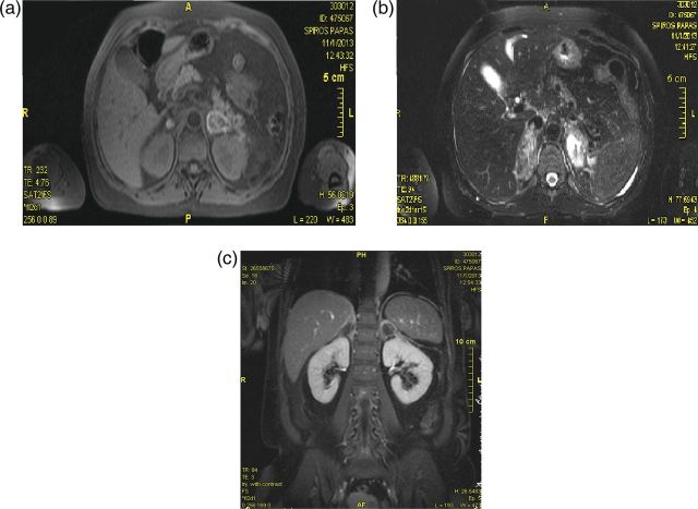
Figure 2:
Magnetic resonance imaging: performed on Day 7 (subacute phase) (a) Transverse view of a T1-weighted image of the adrenals demonstrating high signal in the periphery (b) Transverse plane of a T2-weighted image showing high signal intensity in the adrenals particularly on the left side (c) Coronal view of a contrast-enhanced image displaying heterogeneous hyperintensity without contrast uptake, excluding metastatic infiltration.

