Figure 7.

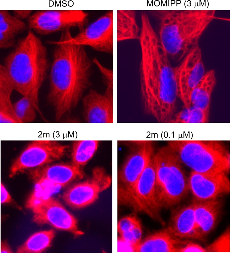
Immunofluorescence imaging of tubulin (red fluorescence) in cells treated for 24 h with the methuosis-inducing compound 1a and the 2-indolyl propyl ester 2m. The nuclei are visualized with DAPI (blue fluorescence).

Immunofluorescence imaging of tubulin (red fluorescence) in cells treated for 24 h with the methuosis-inducing compound 1a and the 2-indolyl propyl ester 2m. The nuclei are visualized with DAPI (blue fluorescence).