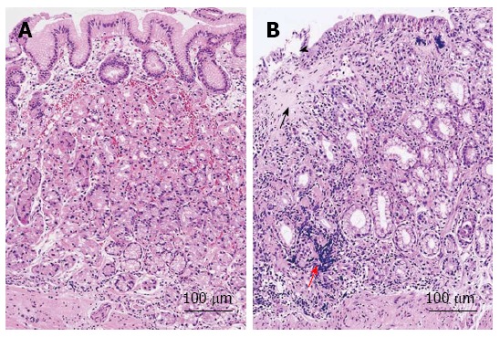
Figure 2.

Histological findings of collagenous gastritis. A: Nodular mucosal lesion did not show marked inflammatory infiltration and collagen deposition; B: Depressive mucosal lesion showed a thick collagen deposition (black arrow) and inflammatory infiltrates (red arrow). The glandular atrophy and epithelial damage is marked (black arrowhead)[34].
