Figure 7.
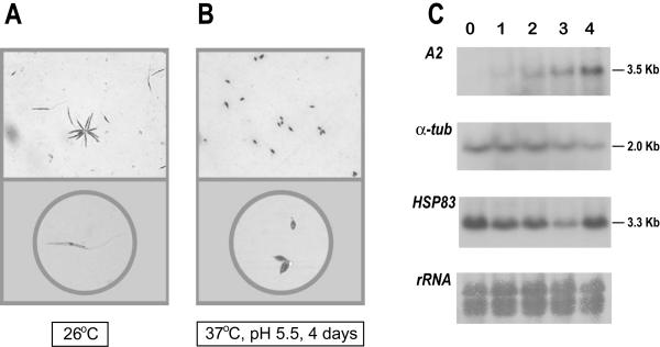
Differentiation of promastigotes to amastigotes in axenic conditions. Morphology of L. infantum promastigotes (A) and amastigote-like forms (B). Logarithmic promastigotes and amastigote-like forms (after 4 days of differentiation) were microscopically evaluated. Parasites were placed on slides and, after air dried, stained with DADE® Diff-Quik® (Baxter Diagnostics, Dudingen, Switzerland). The magnifications used to take the micrographs were: 400 × (top panels) and 630 × (bottom panels). (C) Expression of A2, α-tubulin, and HSP83 transcripts during differentiation of promastigotes to amastigotes. Promastigotes (lane 0) were differentiated to amastigotes (see Materials and methods for details). Aliquots of 5 × 10⁷ cells were taken every 24 h (day 1, 2, 3, and 4) and used to extract RNA. The Northern blot was probed with the L. donovani A2 gene [26] and, after stripping, the blot was reprobed with the T. cruzi α-tubulin gene [38], and the 3'-UTR of the L. infantum HSP83 gene. A methylene blue staining of the blot is shown at the bottom (panel rRNA).

