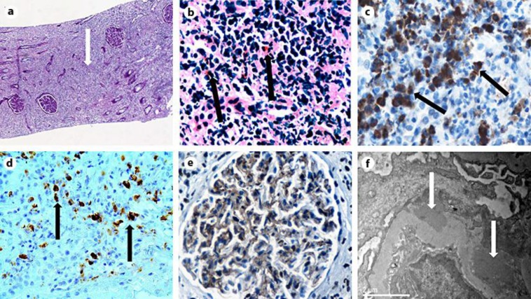
Fig. 1.
a Rarefaction of tubules and glomeruli, severe interstitial inflammatory infiltrates and fibrosis (arrow), PAS ×10. b Interstitial nephritis with dense lymphoplasmacytic infiltrations and eosinophils (arrows), PAS ×400. c The majority of interstitial inflammatory infiltrates were IgG-producing plasma cells (arrows). Immunohistochemistry for IgG, ×400. d The majority (>40%) of IgG+ plasma cells appear positive for IgG4 (arrows). In absolute numbers, >50 IgG4+ plasma cells are seen per hpf. Immunohistochemistry for IgG4, ×400. e Focal thickening of GBMs with C4d positivity indicating MN lesions. Immunohistochemistry for C4d, ×200. f Electron microscopy revealed the presence of subepithelial (arrows) and scarce mesangial deposits compatible with secondary MN, ×15.000.

