
Figure 2.
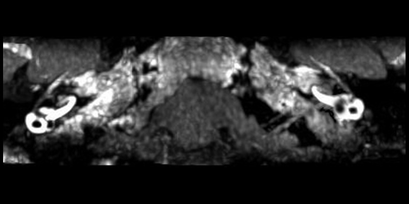
MIP of 3D-FLAIR MRI of patient no. 20. Maximum Intensity Projection (MIP) of original 3D-FLAIR images. The vestibular part of the perilymph is partially enhanced by contrast agent in the right affected side. The cochlea showed no hydrops. Mild hydrops in the vestibular and cochlea can be observed on the left side, which indicates the “asymptomatic” ear is involved.