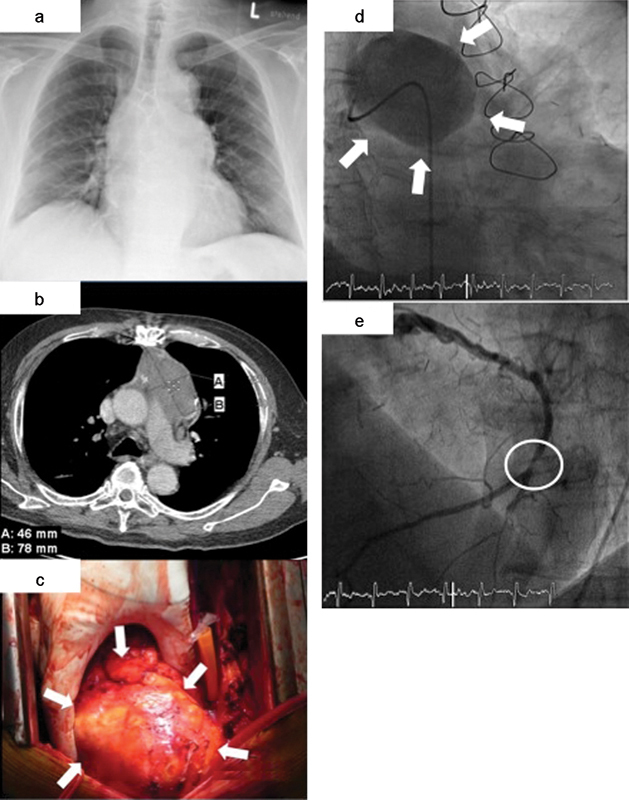
Fig. 1.

(a) Chest X-ray (posteroanterior) with an enlargement of the mediastinum. (b) Venous graft to marginal branches of the circumflex artery. (c) White arrows show the dimension of the giant aneurysm, which lays directly beneath the sternum. (d) Perfused saphenous vein graft aneurysm to the circumflex artery (CX) in the angiography. (e) Distal stenosis of the large CX.
