Figure 1.
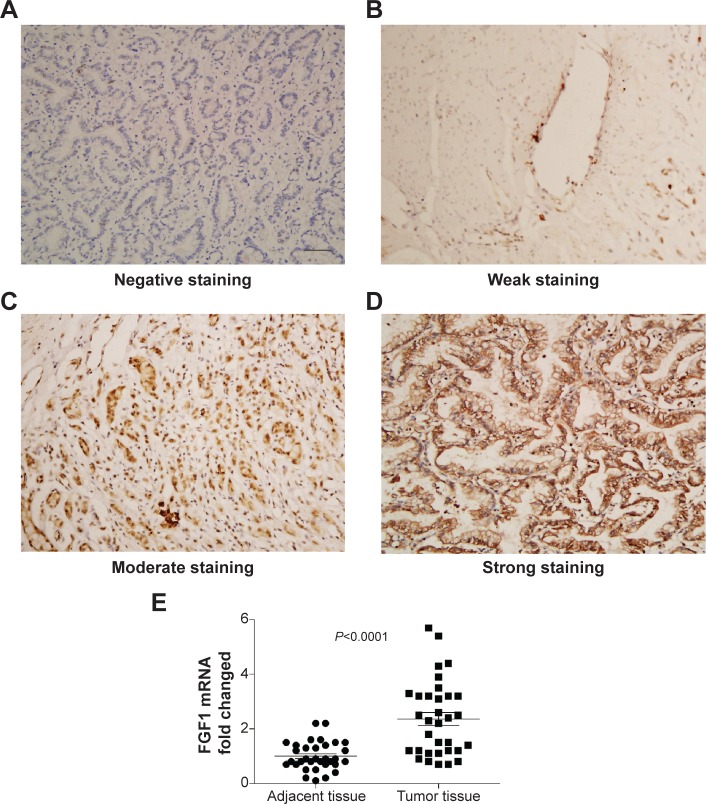
Representative immunohistochemical staining of FGF1 in gastric adenocarcinoma.
Notes: (A) Negative FGF1 staining; (B) weak FGF1 staining; (C) moderate FGF1 staining; (D) strong FGF1 staining; scale bar: 50 μm. (E) The mRNA of FGF1 from tumor tissue and corresponding adjacent tissue was detected by qPCR.
Abbreviations: FGF1, fibroblast growth factor 1; qPCR, quantitative polymerase chain reaction.

