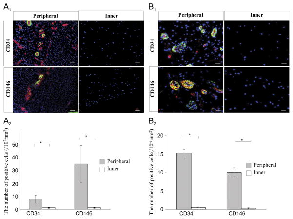
FIGURE 1.
A, Immunohistochemical staining for CD34 and CD146 in fetal meniscal tissues in vivo. A1, The left panels are representative images of the peripheral vascular region, and the right panels are the inner avascular region. CD34, CD146 (red), and α-SMA (green) (scale bars = 25 μm). A2, The number of positive cells for CD34 and CD146 in the fetal meniscus is shown in the graph (*P < 0.05). B, Immunohistochemical staining for the CD34 and CD146 in adult meniscal tissues. B1, The left panels are the peripheral vascular region, and the right panels are the inner avascular region. CD34, CD146 (red), α-SMA (green) (scale bars = 50 μm). B2, The number of positive cells for CD34 and CD146 in the adult menisci are shown in the graph (*P < 0.05). A full color version of this figure can be viewed in the online version of the article (http://journals.lww.com/acsm-msse).

