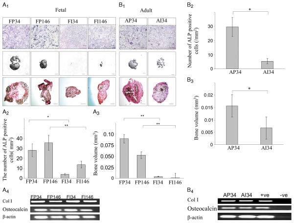
FIGURE 4.
Osteogenic potential of meniscus-derived cells. A, Human fetal meniscus-derived vascular cells were cultured in osteogenic medium. A1, Monolayer culture of fetal meniscus-derived vascular cells from each region of the meniscus were also maintained and treated with osteogenic medium for 21 d (scale bars = 50 μm). Pellet cultures of the meniscus-derived vascular cells from each region of the fetal meniscus were also maintained and treated with osteogenic medium for 21 d. Bone volumes were assessed by micro-CT (scale bars = 250 μm). Pellets were von Kossa stained (scale bars = 250 μm). A2, Quantification of the number of ALP-positive cells is shown in the graph. CD146-positive cells in the peripheral region of the meniscus showed more ALP-positive cells (*P < 0.05). A3, Quantification of bone volume in pellets is shown (*P < 0.05). A4, RT-PCR analysis for mRNA expression of COLI, osteocalcin, and β-actin in the cells of FP34, FP146, FI34, and FI146 after osteogenic induction for 21 d. B. Human adult meniscus-derived vascular cells (CD34-positive cells) were cultured in osteogenic medium. B1, Monolayer culture of meniscus-derived vascular cells from each region of the meniscus were also maintained and treated with osteogenic medium for 21 d (scale bars = 50 μm). Pellet culture of meniscus-derived vascular cells from each region of the meniscus were also maintained and treated with osteogenic medium for 21 d. Bone volumes were assessed by micro-CT (scale bars = 250 μm). Pellets of the adult meniscus-derived cells were von Kossa stained (scale bars = 250 μm). B2, Quantification of the number of ALP-positive cells is shown. CD146-positive cells in the peripheral region of the meniscus showed more ALP-positive cells (*P < 0.05). B3, Quantification of the bone volume of the pellets is shown. (*P < 0.05). (e) Pellets were von Kossa stained (scale bars = 250 μm). B4, RT-PCR analysis for mRNA expression of COLI, osteocalcin, and β-actin in the cells of AP34 and AI34 after osteogenic induction for 21 d.

