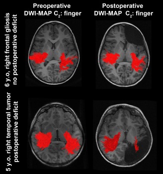
Figure 6.

Representative examples of C2 (finger) pathways (red fibers) obtained from preoperative DWI‐MAP classifier (left column) and postoperative DWI‐MAP classifier (right column). The resection of the presumed epileptogenic zone in the right frontal lobe yielded no loss of the finger pathway (top); in contrast, the resection of the right hemisphere including a tumor resulted in significant loss of the finger pathway (bottom). [Color figure can be viewed in the online issue, which is available at http://wileyonlinelibrary.com.]
