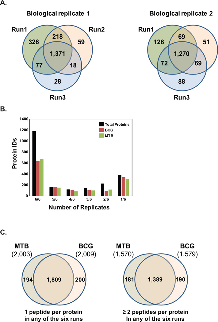
Figure 1. Total proteins identified in the M. tuberculosis (MTB) and M. bovis BCG membrane fractions.
(A) Mass spectrometry analysis of membrane fractions derived from either M. tuberculosis (MTB) or M. bovis BCG (BCG) resulted in the high-level of reproducibility in three technical runs for each of the two biological replicates with biological replicate 1 showing 1,371 identifications in all three technical replicates while biological replicate 2 shows 1,270 identifications in all three technical replicates. (B) Proteins identified in all six runs were categorized on the basis of the number of replicates they were identified, for all proteins in MTB, BCG, or both strains. (C) Protein identifications in MTB and BCG using 1 peptide per protein criteria in any of the six runs and ≥ 2 peptides per protein criteria

