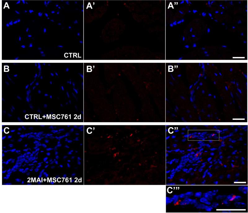
Figure 4. X-Sight-labeled MSCs tracking in the hearts by confocal microscopy.
Hearts of control or chagasic mice treated with labeled MSC were sliced for confocal analysis (A-C’”) Representative confocal images showing X-Sight-labeled cells (red) in heart slices 2 days after therapy. (A-A”) CTRL. (B-B”) CTRL+MSC761 2d. (C-C”) 2MAI+MSC761 2d: (A, B and C) DAPI, (A’, B' and C') X-Sight nanoparticles, and (A”, B” and C”) Merged images. (C”’) Two times magnified image of the white square on (C”). It is possible to note a higher concentration of MSCs in C, which is likely due to extravasation of inflammatory cells or attraction of other cells in the infected heart treated with MSC. Groups abbreviations: CTRL-control mice; 2MAI-2 months after infection; +MSC761 2d-plus X-Sight 761-labeled MSC after 2 days of the transplantation, respectively. Scale bar = 20 μm.

