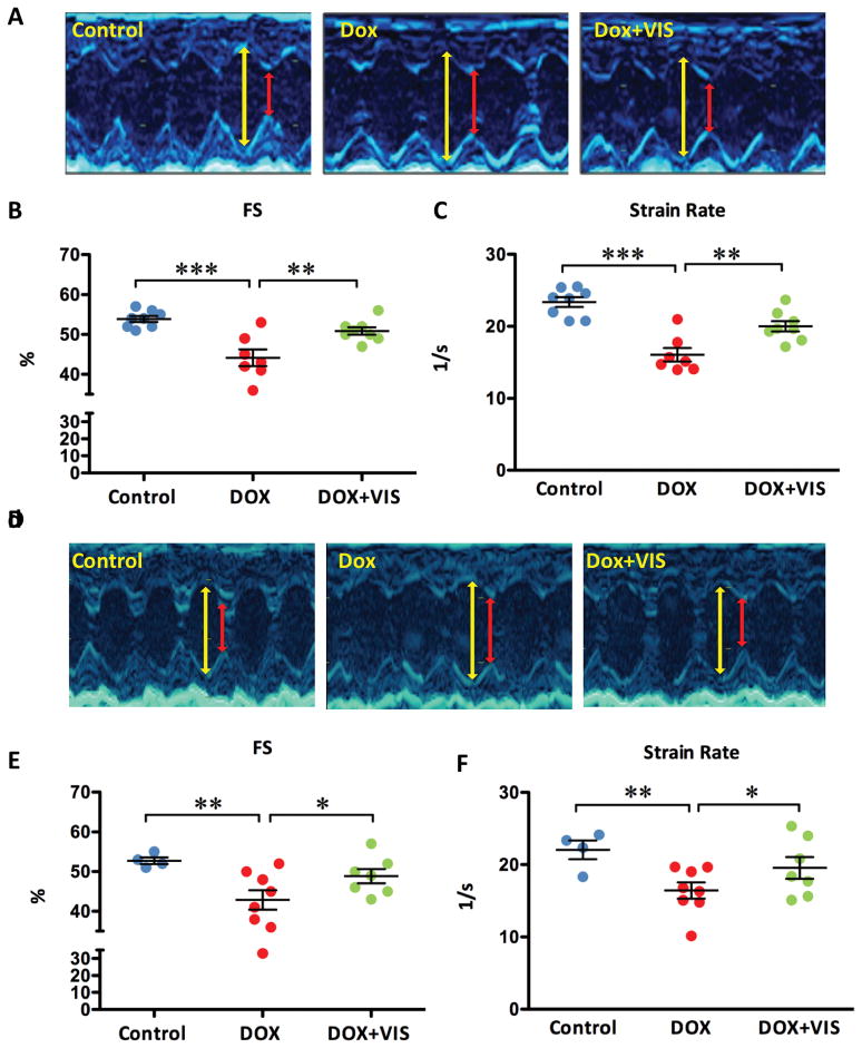
Fig 3. VIS effect on heart function in doxorubicin treated mice.
Representative M-mode echocardiogram images of acute (A) and chronic (D) models: yellow line denotes diastolic left ventricle internal dimension (LVIDd) and red line denotes systolic left ventricle internal dimension (LVIDs). Effects of doxorubicin with and without VIS on fractional shortening (B,E) and strain rate (C,F) in both acute (B,C) and chronic (E,F) models. For acute models (B,C), groups were compared using one-way ANOVA followed by the Tukey post-test. For chronic models (E,F), groups were compared using one-way ANOVA followed by a one-tailed Student’s T-test. Cont – vehicle treated control mice, Dox – doxorubicin treated mice, Dox + VIS – doxorubicin and VIS co-treated mice. Statistics (compared to doxorubicin treated samples): * p< 0.05, ** p<0.01, and *** p<0.001.

