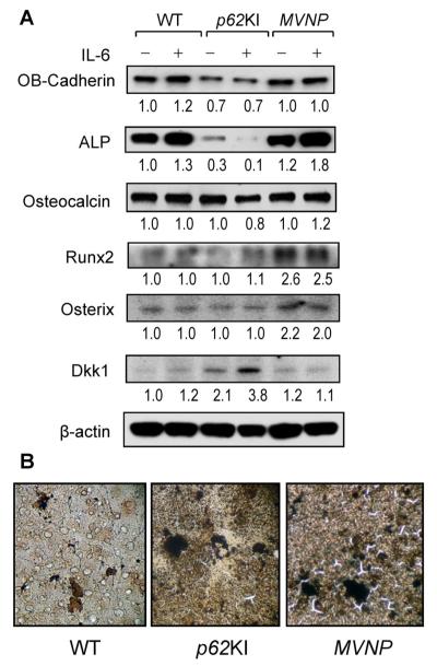
Fig. 6.
Osteoblast differentiation markers in osteoblasts derived from WT, p62KI, and MVNP mice. (A) Expression of osteoblast differentiation markers. Primary osteoblasts (2 × 10⁵ cells/35-mm dish) were cultured with or without 10 ng/mL of IL-6 for 4 days in 10% FCS in α-MEM. Cell lysates were analyzed by immunoblot using antibodies recognizing OB-Cadherin (Cell Signaling, Beverly, MA, USA), alkaline phosphatase (Millipore, Billerica, MA, USA), Runx2 (Santa Cruz Biotechnology, Santa Cruz, CA, USA), Osterix (Abcam, Cambridge, MA, USA), osteocalcin (Millipore), Dkk1 (Cell Signaling), and β-actin (Abcam) as loading control. (B) Calcification in vitro. Osteoblasts were cultured in 10% FCS with α-MEM for 3 weeks as described in Subjects and Methods. The cells were stained with von Kossa stain (×100). WT = wild-type; IL-6 = interleukin 6; FCS = fetal calf serum; α-MEM = α modified essential medium.

