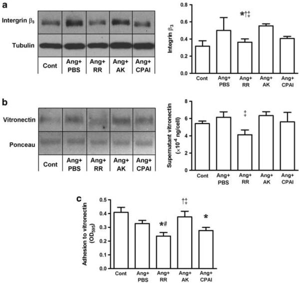
Figure 3.
Changes in integrin β3 expression, supernatant vitronectin and adhesion of cardiac fibroblasts to vitronectin. In cardiac fibroblasts exposed to Ang II, non-vitronectin-binding AK (Ang + AK) significantly upregulated cellular expression of integrin β3 (western blot, a) compared with Ang II alone (Ang + PBS) and the vitronectin-binding PAI-1 variants, RR (Ang + RR) and CPAI (Ang + CPAI). This AK-caused increase in integrin β3 was accompanied by higher level of supernatant vitronectin than with RR treatment (western blot, b) and greater adhesion to vitronectin than those in response to RR and CPAI (c). These results suggest a decreased vitronectin–αVβ3 interaction by vitronectin-bound PAI-1. * vs Cont, # vs Ang + PBS, † vs Ang + CPAI, † vs Ang + RR, P <0.05. N = 3 for each group.

