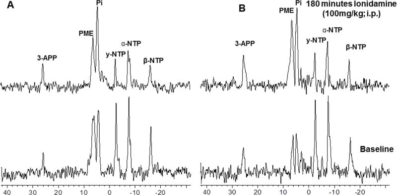
Figure 1.
In vivo localized (Image Selected In vivo Spectroscopy - ISIS) 31Phosphorus magnetic resonance (31P MR) spectra of human melanoma xenograft grown subcutaneously in nude mice (A) without glucose infusion and (B) with glucose (26 mM) infusion. In each set lower spectrum represents baseline and upper represents 180 min following lonidamine (LND; 100 mg/kg, i.p.) administration. Resonance assignments are as follows, 3-APP (3-aminopropylphosphonate); PME (phosphomonoesters); Pi (inorganic phosphate); PDE (phosphodiesters); γ-NTP (γ-nucleoside-triphosphate), α-NTP (α-nucleoside-triphosphate), β-NTP (β-nucleoside-triphosphate). Decrease in β-NTP levels and the corresponding increase in Pi following LND administration (Spectrum B) indicating impaired energy metabolism.

