
Figure 4. SAHA decreases enhanced TNF-α production stimulated by LPS or Pam3CSK4 in culture supernatant of RAW264.7 macrophages.
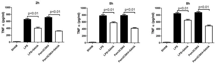
Concentrations of TNF-α and IL-6 in culture supernatant of RAW264.7 Macrophages were determined by ELISA at 2, 5, and 8 h after LPS or Pam3CSK4 treatment in the absence or presence of SAHA. Untreated macrophages served as control (means ± SEM, n = 3).