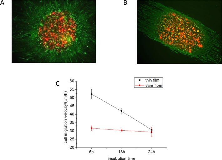
Fig 1. En-mass cell migration within 24 hours.
The overlapped image of en-mass cell migration of live cells stained with DiD (red), and incubated for four hours, onto the image of cells incubated for 24h, fixed and stained for F-actin with Alexa Fluor (green) (a) On a spun cast, FN coated PMMA thin film and (b) On electrospun FN coated, PMMA microfibers. The lines are drawn to guide the eye towards the perimeter of the migration front at 4 and 24 hours, respectively. Error Bar = 250 μm. (c) The en-mass cell migration velocity, as measured from the motion of the front, on the thin film (black) and 8μm fibers (red) as a function of time.

