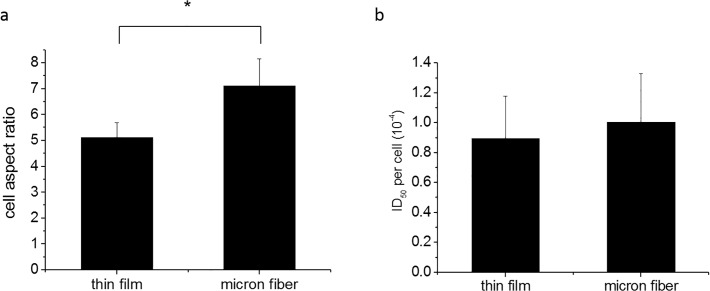
Fig 3. Cell aspect ratio and ID50 per cell reading on thin film and microfibers at day 4.
(a) Cell aspect ratio was calculated as: the length of the cell/width of the cell plated on thin film and micron fiber surfaces, P<0.001 (*). (b) The XTT assay at day 4 was performed and followed with a DAPI nuclear staining. The reading of ID50 value reflects the metabolism level of all the cells at day 4. However, the cell proliferation varied on different surfaces. As a result, we divided the ID50 value by the actual nuclear number counted after DAPI staining, and evaluate the metabolism level per cell. P>0.05 which was not considered as significant.

