
Fig 4. Infection of MDMs by X31.
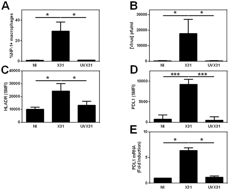
MDM were differentiated in the presence of 2 ng/ml GM-CSF for 12 d prior to infection with 500 pfu/ml of H3N2 X31 influenza virus or a UV-irradiated aliquot of virus (UVX31) for 2 h. After washing, media was replaced and the cells incubated for a further 22 h before supernatants and cells were harvested. Cells were analysed using flow cytometry and RT-PCR, supernatant was analysed using dot blot. Histograms showing infected MDM expression of A) Viral NP1 expression (% cells n = 7), B) release of hemagglutinin into supernatants (n = 10) C) cell surface HLA-DR expression (specific mean fluorescence intensity—SMFI n = 7) D), PDL1 expression (SMFI n = 7) E) PDL1 gene expression (n = 6). Data are expressed as means ± SE of n independent experiments and analysed using a paired t-test * p<0.05, ** p<0.01. *** p<0.001.