
Fig 5. Functional effect of PDL1 on T cell activation.
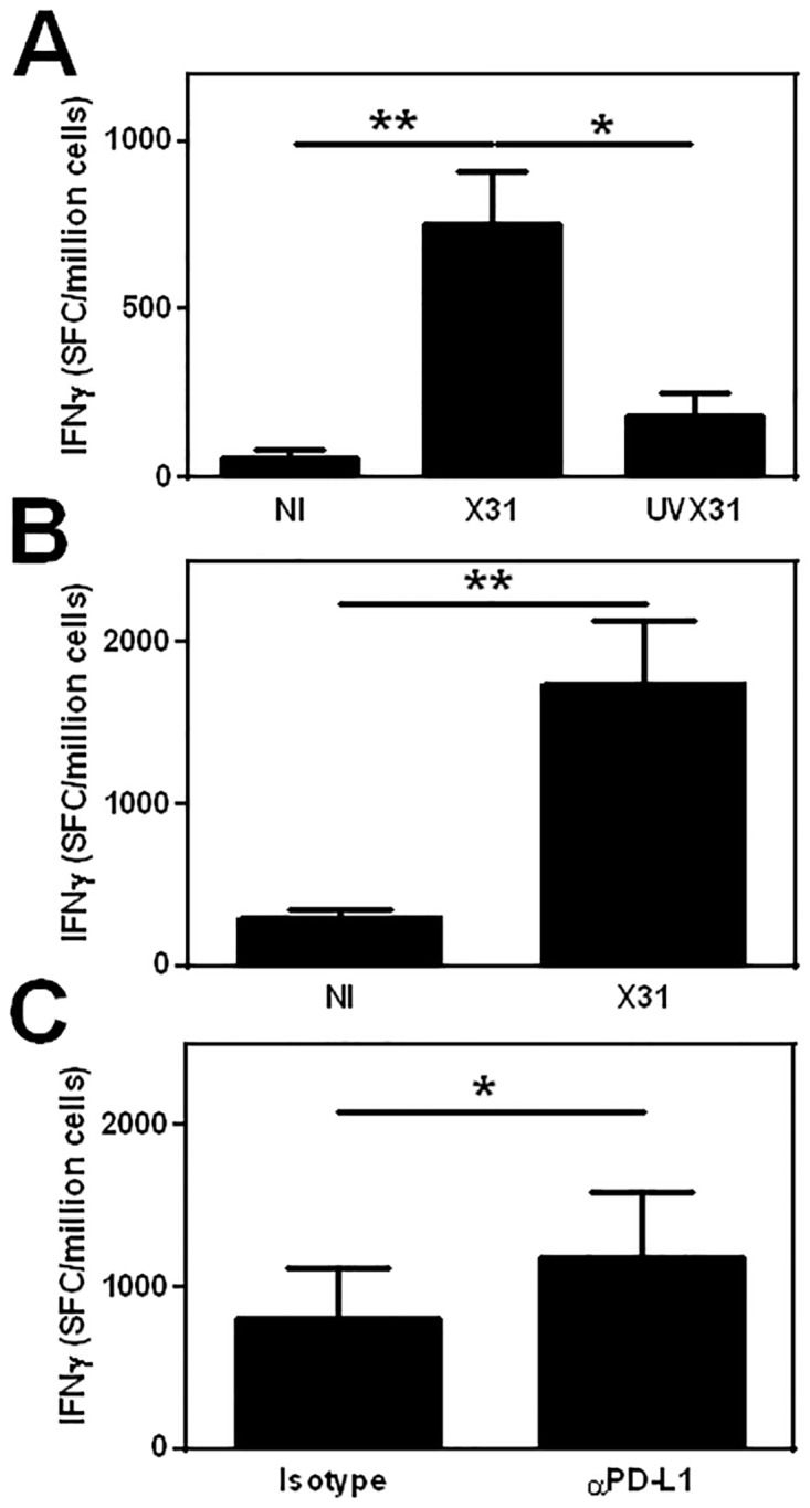
MDM were differentiated in the presence of 2 ng/ml GM-CSF for 12 d prior to infection with 500 pfu/ml of H3N2 X31 influenza virus or a UV-irradiated aliquot of virus (UVX31) for 2 h. Infected MDM were transferred to a coated ELISpot plate and autologous lymphocytes added and incubated for a further 22 h before IFNγ release was measured using ELISpot. A) 2.5 x 10⁵ monocyte-depleted PBMC co-cultured with 5 x 10⁴ autologous MDM not infected (NI) or treated with X31 or UVX31(n = 5). B) 1 x 10⁵ CD8+ T cells co-cultured with 5 x 10⁴ autologous MDM NI or infected with X31 (n = 6) C) 1 x 10⁵ CD8+ T cells co-cultured with 5 x 10⁴ autologous MDM infected with X31 in the presence of 10 μg/ml anti-PDL1 antibody or isotype control (n = 5). Data are expressed as means ±SE of n independent experiments and analysed using a paired t-test * p<0.05, ** p<0.01.