Figure 1.

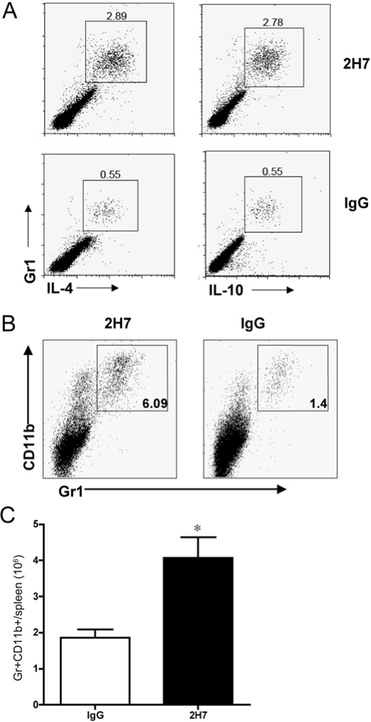
B cell depletion induced regulatory Gr1+ cells that express IL-4 and IL-10. A) Splenocytes from anti-CD20 (2H7) or control IgG treated hCD20.NOD mice (n=5/group) were stimulated with 50 ng/ml PMA and 500 ng/ml ionomycin for 5 hours in the presence of GolgiStop (BD) and then stained with monoclonal antibodies against the surface markers CD4, CD11b and Gr1, followed by intracellular staining with anti-IL-4 and anti-IL-10 or isotype control antibody. One representative set of FACS plots is shown in Figure 1A with gating on CD4− cells. B) Splenocytes from mice treated with 2H7 or control mouse IgG were stained with monoclonal antibodies against the surface markers Gr1 and CD11b. C) Gr1+CD11b+ cell number was enumerated in splenocytes from 2H7 or control mouse IgG treated mice (n=4 each group, p = 0.011, Student’s t test).
