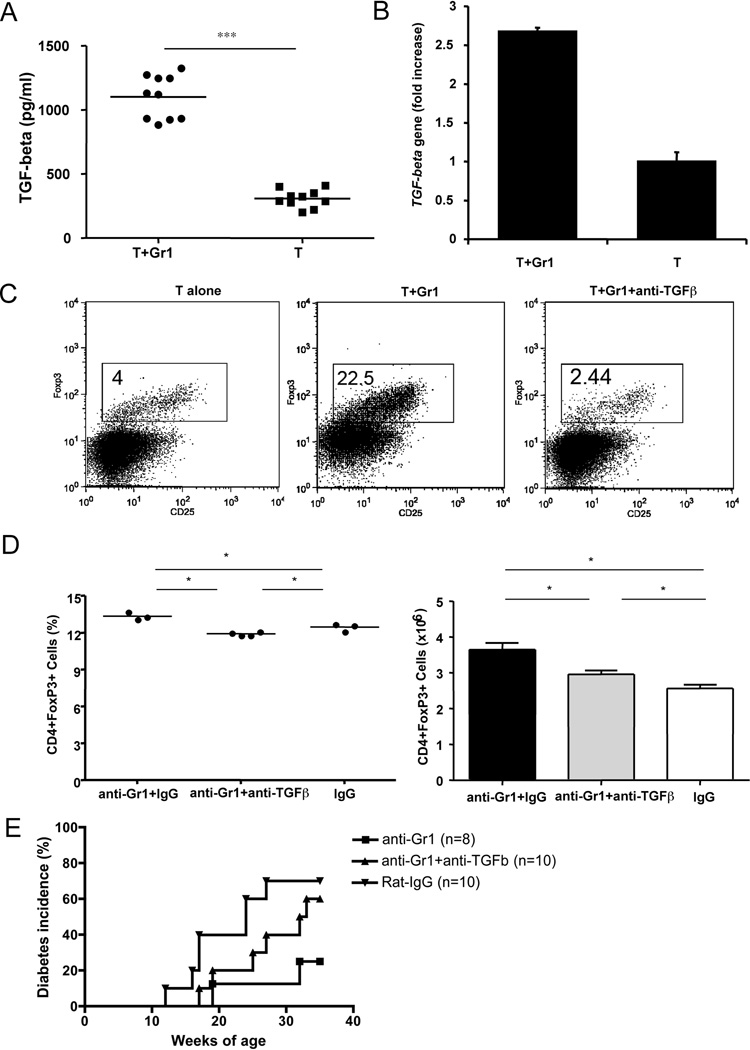
Figure 8.
Gr1+CD11b+ cells induce regulatory T cell differentiation in a TGF-beta dependent manner. A) Secreted TGF-beta from culture supernatants (Fig. 7C) was tested by ELISA (BD Bioscience). Purified naïve T cells were co-cultured with purified Gr1+CD11b+ cells (T+Gr1) compared with T cells alone (T). *** p<0.0001. B) TGF-β expression was detected at transcriptional level by qPCR (5’-primer: TGGAGCCTGGACACACAGTA, 3’-primer: GTTGGACAACTGCTCCACCT). qPCR was performed in triplicate in 3 separate experiments. Data from one of 3 experiments are shown. *** p<0.0001. C) Purified Gr1+CD11b+ cells were co-cultured with purified naïve T cells at 1:5 ratio for 5 days in the presence or absence of neutralizing anti-TGF-β (clone 1D11; 10 µg/ml). The differentiation of naïve T cells into regulatory T cells was determined by flow cytometry. Three independent experiments were performed. D) and E) Nine week-old female NOD mice were injected with one dose of 0.25mg anti-Gr1 or control IgG antibody intravenously together with or without neutralizing anti-TGF-β (3 injections of 0.25 mg/mouse, 3 day intervals). FoxP3+CD4+ cells were examined at 35 weeks of age in both antibody and control IgG treated mice (D, percentage on the left and absolute numbers on the right, *p<0.05), together with the development of diabetes (E). Diabetes development was monitored by testing for glycosuria twice a week until 35 weeks of age. Disease was confirmed by a blood glucose level greater than 250 mg/dl. P=0.035 for anti-Gr1 vs anti-Gr1+anti-TGFβ groups; p=0.331 for control IgG vs anti-Gr1+anti-TGFβ groups; p=0.045 for anti-Gr1 vs control IgG groups.

