FIGURE 4.
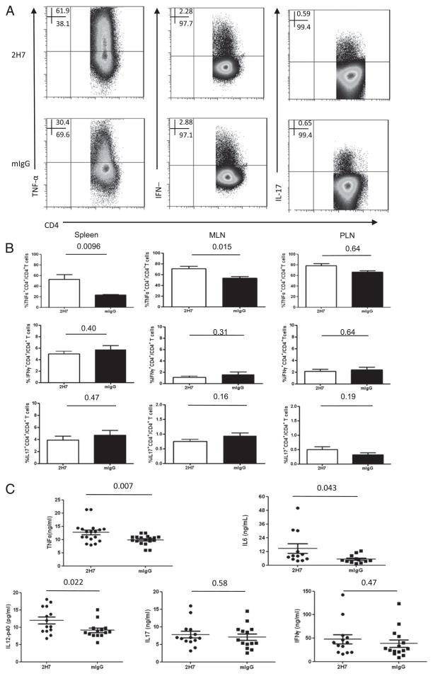
BDC2.5 T cells produce more inflammatory cytokines after B cell depletion. HCD20-BDC2.5NOD mice were sacrificed after 2H7 or mIgG treatment. Spleen, MLN, and PLN cells were stimulated with BDC2.5 mimotope (10 ng/ml) overnight, followed by intracellular staining for cytokines. (A) Representative dot plots for inflammatory cytokine production by splenic BDC2.5 T cells after 2H7 and mIgG treatment. (B) Summary of percentage of inflammatory cytokine-producing BDC2.5 T cells (TNF-α+CD4+, IFN-γ+CD4+, and IL-17+CD4+) on gated CD4+ T cells (mean ± SEM; n = 4) in B cell-depleted (□) or control IgG (■)-treated hCD20-BDC2.5NOD mice. Similar results were obtained from three independent experiments. (C) Circulating plasma cytokines including TNF-α, IL-6, IL-12p40, IL-17, and IFN-γ were measured by Luminex assay from B cell-depleted and control IgG-treated mice (n > 12/group). All the numbers above the lines in (B) and (C) are p values.

