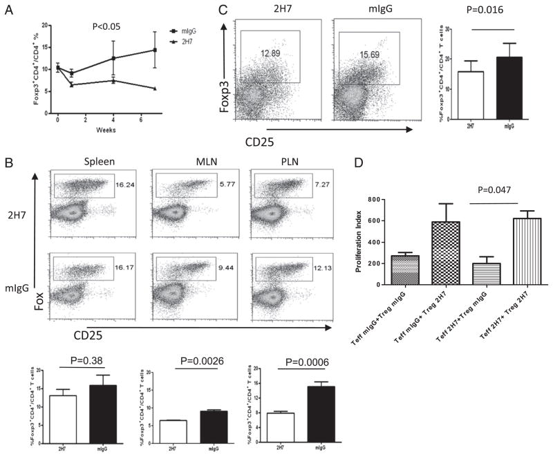
FIGURE 5.
B cell depletion reduces BDC2.5 Treg number and function. (A) Foxp3+ Treg cells from PBMC were analyzed by flow cytometry following treatment of hCD20-BDC2.5NOD mice with 2H7 or mIgG. The percentage of BDC2.5 Treg cells decreased in 2H7-treated mice (n = 6–8 mice/group; p < 0.05). (B) Percentages of Treg in BDC2.5 T cells in different lymphoid organs were variable. Representative dot plots show the percentage of BDC2.5 Treg (Foxp3+CD25high+low) cells. The bar charts show the pooled data from at least six mice per group (mean ± SEM). (C) BDC2.5 Tregs in islets from 2H7- or mIgG-treated hCD20-BDC2.5NOD mice. After islet isolation, the islet cells were dispersed, and infiltrating cells were stained with the corresponding mAbs and analyzed by flow cytometry. There were fewer infiltrating Treg cells in islets after B cell depletion. Dot plots are shown after gating on CD4+ cells. At least two mice were pooled for islet isolation. Experiments were performed three times and summarized in the bar graphs on the right. (D) Cross-suppression assay. Foxp3+ BDC2.5 Treg cells were sorted from 2H7- or mIgG-treated hCD20-BDC2.5-FirNOD mice and used as Treg (Foxp3+) and Teff cells (Fir−CD25−CD4+) were also sorted from 2H7- or mIgG-treated hCD20-BDC2.5-FirNOD mice. As indicated in (D), the cross-suppression assay was performed by coculturing Teff cells from 2H7-treated mice with Treg cells from mouse IgG-treated mice or Teff cells from IgG-treated mice with Treg cells from 2H7-treated mice. Cocultures of 2H7-treated Teff and Treg cells or IgG-treated Teff and Treg cells were used as controls. A cell ratio of 2:1 (Teff/Treg) was used in the cocultures. Proliferation is shown as stimulation index. Treg cells from 2H7-treated mice do not suppress Teff cell proliferation as effectively as Treg cells from mIgG-treated mice. The average [3H]thymidine incorporation in the absence of mimotope = 74.65 ± 13.48 cpm. Similar results were obtained from three independent experiments.

