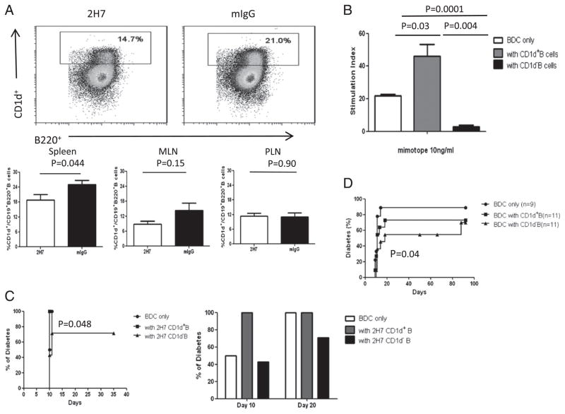
FIGURE 7.
CD1d-B cells are more protective than CD1d+ B cells. HCD20-BDC2.5NOD mice were sacrificed 2 mo after the last treatment. (A) Representative dot plots (top panels) showing regenerated B cells (gated on CD19+) from 2H7- or mIgG-treated hCD20-BDC2.5NOD mice as determined by inmmunofluorescent staining with CD1d, CD19, and B220. At least three mice per group were used for each experiment, and the experiments were performed three times as summarized in the lower panels. (B) Purified BDC2.5 T cells were cocultured with three times the number of sorted CD1d+ B cells (CD19+B220+) or CD1d− B cells (irradiated after sorting) in the presence of 10 ng/ml mimotope. Stimulation index was calculated as described earlier. Background cpm in the absence of mimotope was 80.17 ± 7.1. p = 0.0012 for three groups overall. Similar results were obtained from three independent experiments. (C) Purified BDC2.5 T cells (0.5 × 10⁶) were cotransferred with sorted CD1d+ or CD1d− B cells (1.5 × 10⁶) taken from 2H7-treated hCD20-BDC2.5NOD mice in the B cell regeneration phase (2 mo after the treatment). Mice were screened daily for diabetes. Mice transferred with BDC2.5 T cells only (n = 6) were compared with mice transferred with BDC2.5 T cells together with CD1d+ 2H7-treated B cells (n = 10); p = 0.08. Mice transferred with BDC2.5 T cells together with CD1d+ 2H7-treated B cells were compared with mice transferred with BDC2.5 T cells together with CD1d- 2H7 B cells (n = 10); p = 0.048. (D) Purified BDC2.5 T cells (106) with sorted CD1d+CD19+B220+ B cells or CD1d−CD19+B220+ B cells from 6-wk-old NOD mice were transferred (3 × 10⁶) into NOD.SCID recipient mice. For comparison between BDC2.5 T cells with or without CD1d-B cells, p = 0.04; for comparison of BDC2.5 T cells with or without CD1d+B cells, p = 0.88.

