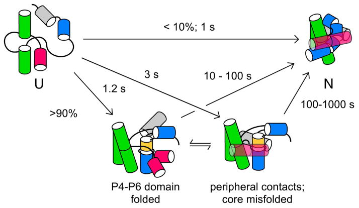
Figure 2. Folding pathway of the Tetrahymena ribozyme.
Time-resolved footprinting30,31 showed that tertiary interactions in the P4–P6 domain (green) form in ~1 s, more rapidly than contacts in peripheral helices (pink and grey) and in the P3–P9 domain (blue), due to mispairing of the P3 helix (gold).24 The ensemble of unfolded structures (U) partitions among parallel pathways, with 5–10% of molecules folding directly to the native state (N).25

