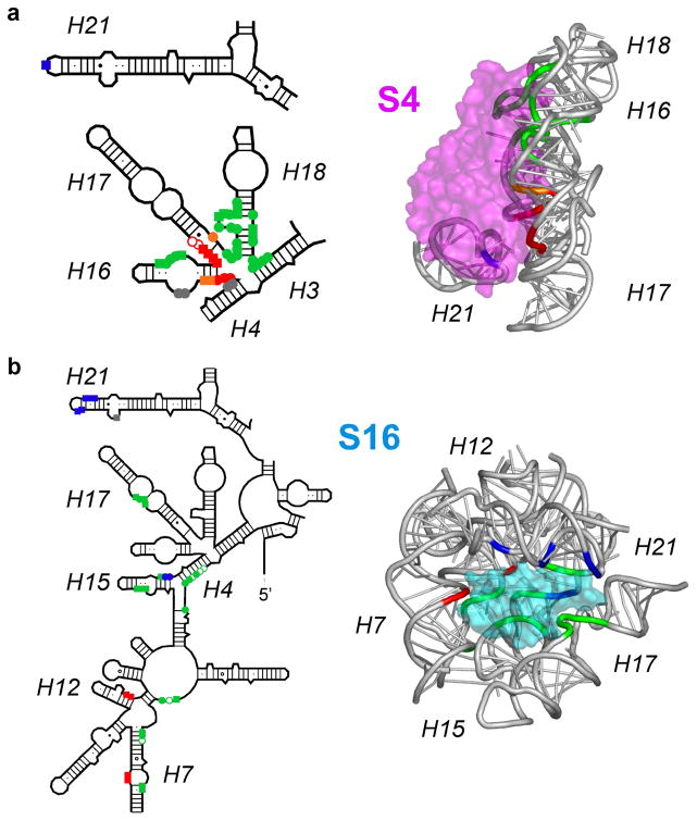
Figure 4. Induced fit in rRNA-protein interactions.
Different residues in a single 30S protein binding site are protected with different rate constants, when probed by X-ray footprinting. (a) Residues contacted by protein S4 in mature 30S subunits colored according to the rate of protection: red, ≥ 20s−1; orange, 2–20 s−1; green, 0.2 –2 s−1; blue, 0.01 – 0.2 s−1. (b) Residues contacted by protein S16, colored as in (a). Reproduced from Ref. 55 with permission.

