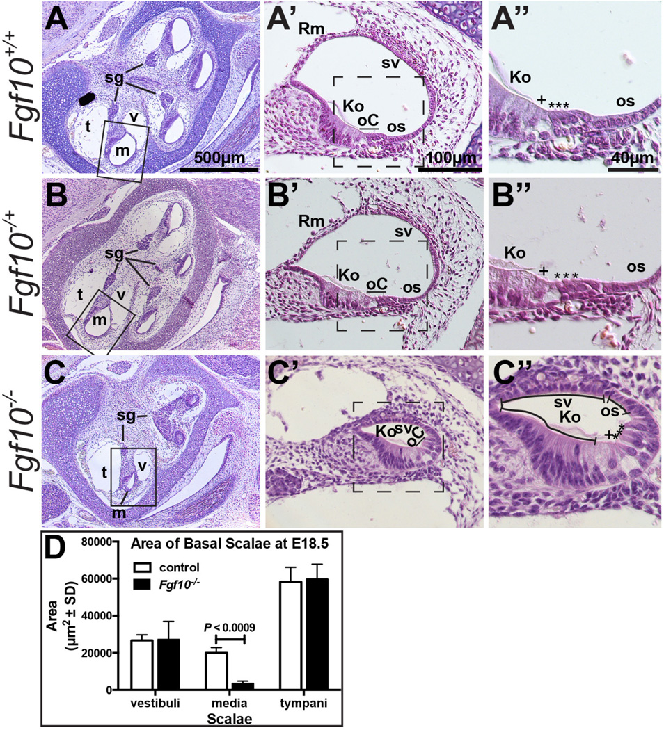
Figure 2. Fgf10 null cochlear ducts lack non-sensory domains and have a reduced cross-sectional area.
Hematoxylin and eosin-stained E18.5 cochlear duct cross sections at three magnifications. Boxes in A–C indicate the region magnified in A’–C’. Dashed boxes in A’–C’; indicate the region magnified in A”–C”. C” Morphologic structures remaining in Fgf10 mutants are indicated with lines. Genotypes are indicated to the left of each row. D. Graphical comparison of the cross sectional area of basal scalae (n = 3 controls and 3 mutants). Abbreviations: Ko, Kolliker’s organ; m, scala media (cochlear duct), oC, organ of Corti; os, outer sulcus; Rm, Reissner’s membrane; sg, spiral (cochlear) ganglion; sv, stria vascularis; t, scala tympani; v, scala vestibuli. Asterisks indicate outer hair cells, plus symbols indicate inner hair cells. Scale bars in A, A’, A’’ apply to all panels in the same column.

