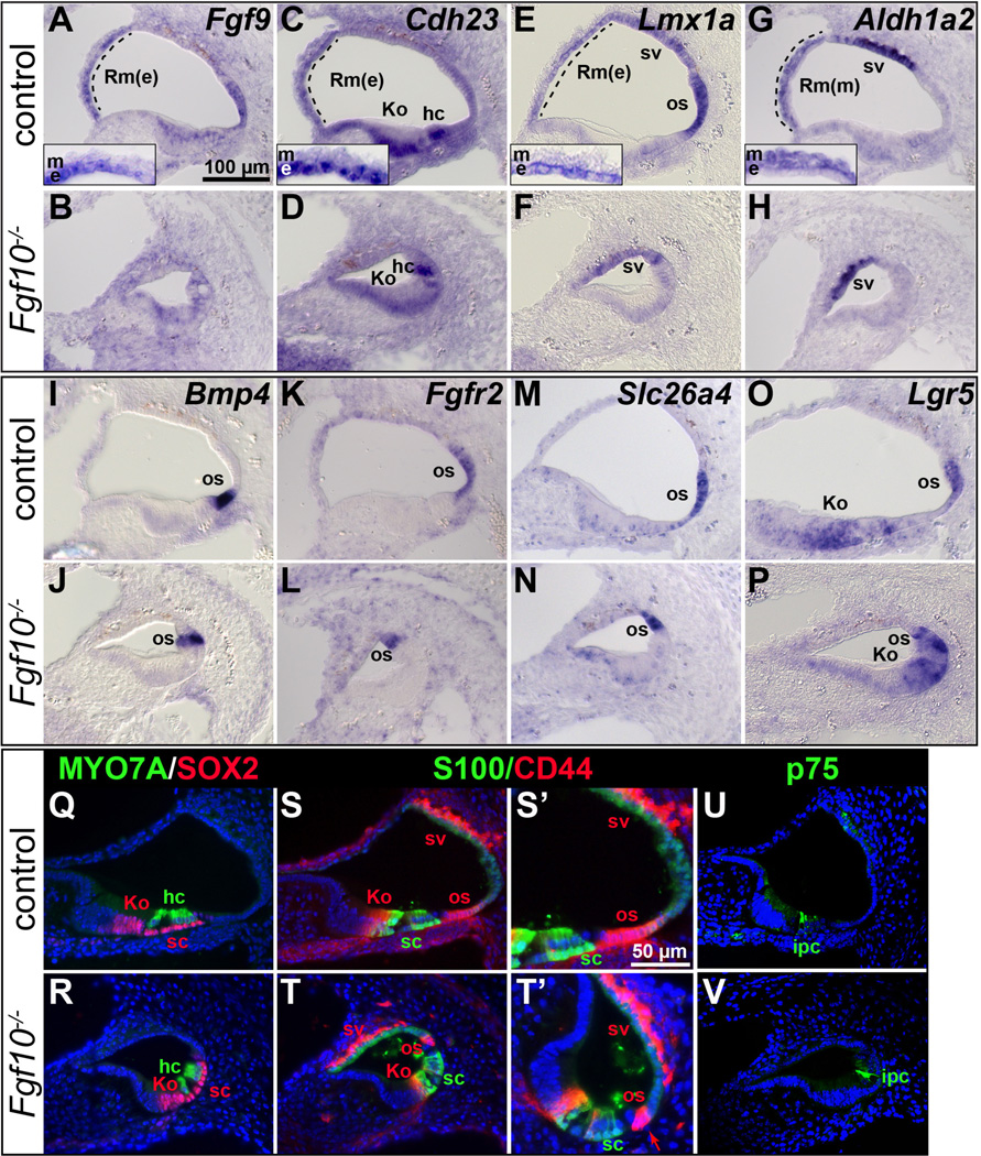
Figure 4. Markers of Reissner’s membrane are absent and outer sulcus markers show a reduced domain in E18.5 Fgf10 null mutant cochleae.
In situ hybridization (A–P) and immunostaining (Q–V) analyses of basal cochlear duct cross sections. Genotypes are indicated to the left of each row and probes are indicated to the upper right of each pair of panels. Insets in A, C, E, G show magnifications of Reissner’s membrane. S’–T’ provide enlargements of S–T, with the arrow in T’ indicating the remnant Claudius cells in the mutant os. Abbreviations: Rm(e), Reissner’s membrane-epithelial layer; Rm(m), Reissner’s membrane-mesenchymal/mesothelial domain; os, outer sulcus. See Figure 2 or 3 legend for others. Scale bar in A applies to all panels except S’–T’. Scale bar in S’ applies to T’.

