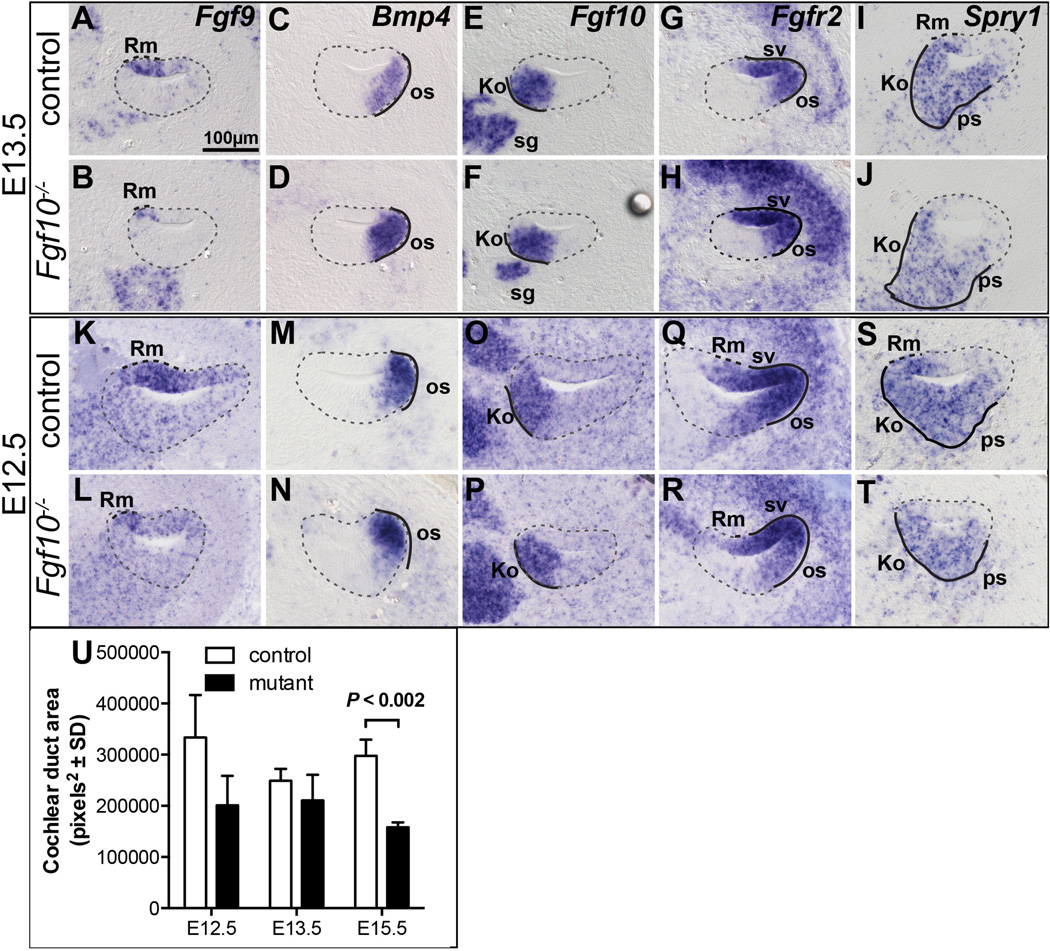
Figure 6. The effects of FGF10 absence on Reissner’s membrane development precede those on outer sulcus development.
In situ hybridization analyses of basal cochlear duct cross sections at E13.5 (A–J) and E12.5 (K–T). Genotypes are indicated to the left of each row and probes are indicated to the upper right of the top panels. Dashed and solid black lines indicate expression that is altered or unchanged, respectively, in mutants. The cochlear duct is outlined with a dashed grey line. U. Graphical comparison of the cochlear duct area of the basal scala media at the developmental ages shown (n = 3 controls and 3 mutants). Abbreviations: Ko, Kolliker’s organ; Rm, presumptive Reissner’s membrane; os, outer sulcus; ps, prosensory region; sg, spiral ganglion; sv, stria vascularis. Scale bar in A applies to all panels.

