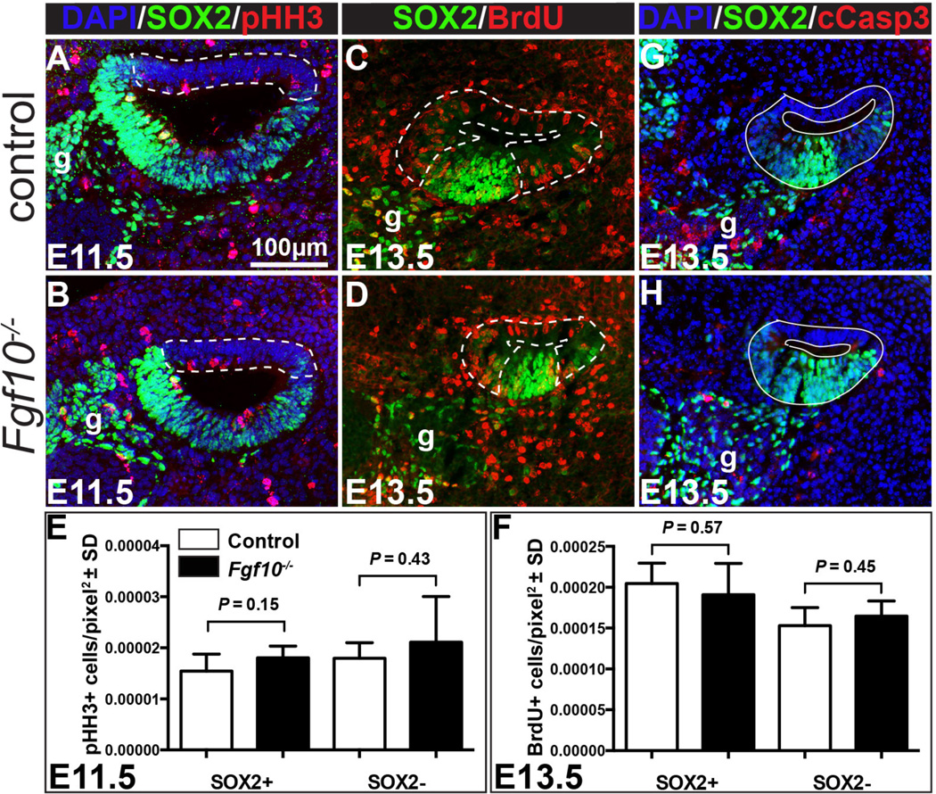
Figure 7. Neither cell proliferation nor cell survival is altered in Fgf10 null cochlear ducts.
Double labeling of control (A) and Fgf10 null (B) E11.5 cochlear duct cross sections with antibodies directed against phospho-Histone H3 (red) and SOX2 (green). Nuclei are counterstained with DAPI (blue). Double labeling of control (C) and Fgf10 null (D) E13.5 cochlear duct cross sections with antibodies directed against BrdU (red) and SOX2 (green). Dotted lines delineate the regions considered non-sensory (SOX2−). The remaining SOX2+ areas were considered prosensory. Graphical comparisons of the mean number of proliferating cells per pixel2 in SOX2+ (E) and SOX2− (F) regions of control (white bars) and Fgf10 null mutants (black bars). Error bars indicate standard deviation (SD). Double labeling of control (G) and Fgf10 null (H) E13.5 cochlear duct cross sections with antibodies directed against cleaved Caspase 3 (cCasp3; red) and SOX2 (green). Nuclei are counterstained with DAPI (blue). Abbreviation: g, ganglion. Scale bar in panel A applies to all images.

