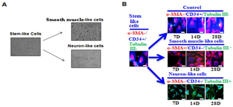
Fig. 6. EC ID3+ stem-like cells display pluripotent morphology.

(A) EC ID3+ stem-like cells were differentiated under defined SMC or neuronal media. Images are from phase-contrast microscopy. EC ID3+ were cultured as spheroids for 7d followed by 7d monolayer culture with differentiation medium. For SMC-like morphology spheroids were cultured with DMEM-F12 containing SMGS (Gibco). For neuron-like cells spheroids were cultured with Neurobasal® Medium (Gibco), StemPro® Neural Supplement (Gibco) and NGF-7s (Sigma). Magnification X 200. (B) Cell differentiation markers were determined using immunofluorescence at days 7, 14, and 28. For immunofluorescence imaging studies, slides were incubated with primary antibodies anti-CD34, α-smooth muscle actin (α-SMA), and β-tubulin III. Nuclei were counterstained with DRAQ5®. Scale bar=20μm. Images were captured with DeltaVision ELITE Olympus IX71 fluorescence microscope, Applied precision (Thermo Scientific) using software softWoRx-Acquire Version: 5.5.1 Release 3.
