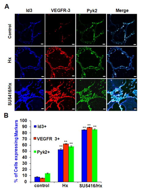
Fig. 8. In vivo pulmonary vascular lesions overexpress ID3, VEGFR3, and Pyk2.
In vivo pulmonary vascular lesions overexpress ID3, VEGFR3, and Pyk2. Lung vascular lesions from the rodent SuHx model of severe PAH were stained with antibodies against: ID3 (blue), VEGFR3 (red), and Pyk2 (green) followed by immunofluorescent detection by confocal microscopy. (A) Both vehicle control and 3 wk chronic hypoxia treatment groups showed a uniform monolayer of cells lining the lumen of the pulmonary artery. SU5416-treated lungs exposed to chronic hypoxia for 3 wk (bottom row) showed a partially blocked lumen with cells stacked on top of each other instead of forming a monolayer. Representative images are from one of three lungs per group. Scale bar = 70 μm (B) Percent of cells expressing each marker amongst treatment groups when treated with vehicle control, hypoxia only, and SU5416 + hypoxia. Error bars represent the mean of expressed marker positive cells ± SD. ** p<0.01 vs. Control in normal/control (810), exposed in hypoxia and Sugen 5416/chronic hypoxia condition of lung tissue. Data were analyzed by ANOVA; Tukey HSD test for multiple comparisons.

