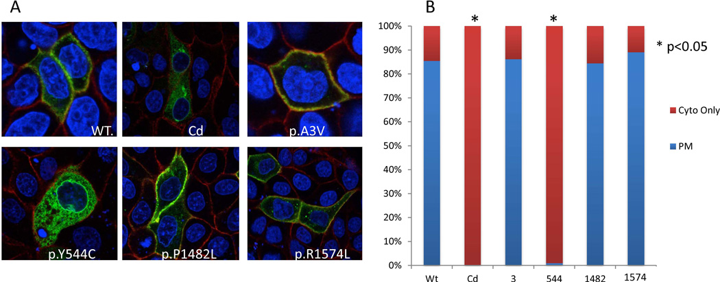
Figure 3.
Subcellular localization of LRP6 variants. (A) Protein subcellular localization in MDCK II cells. Blue color indicates DAPI stain of the nucleus, red color indicates SCRIB stain for plasma membrane, and green color indicates GFP from the LRP6-GFP fusion plasmid constructs. (B) Quantitative analysis of protein localization for GFP-Lrp6 wildtype and novel variants. Fields of cells were imaged randomly and at least 100 GFP positive cells were counted per construct for each of the triplicates. Cells were classified as either GFP only in the cytoplasm or GFP predominantly in the plasma membrane. Asterisks represent a significant (p<0.05) departure from the wildtype construct using a chi-squared analysis.

