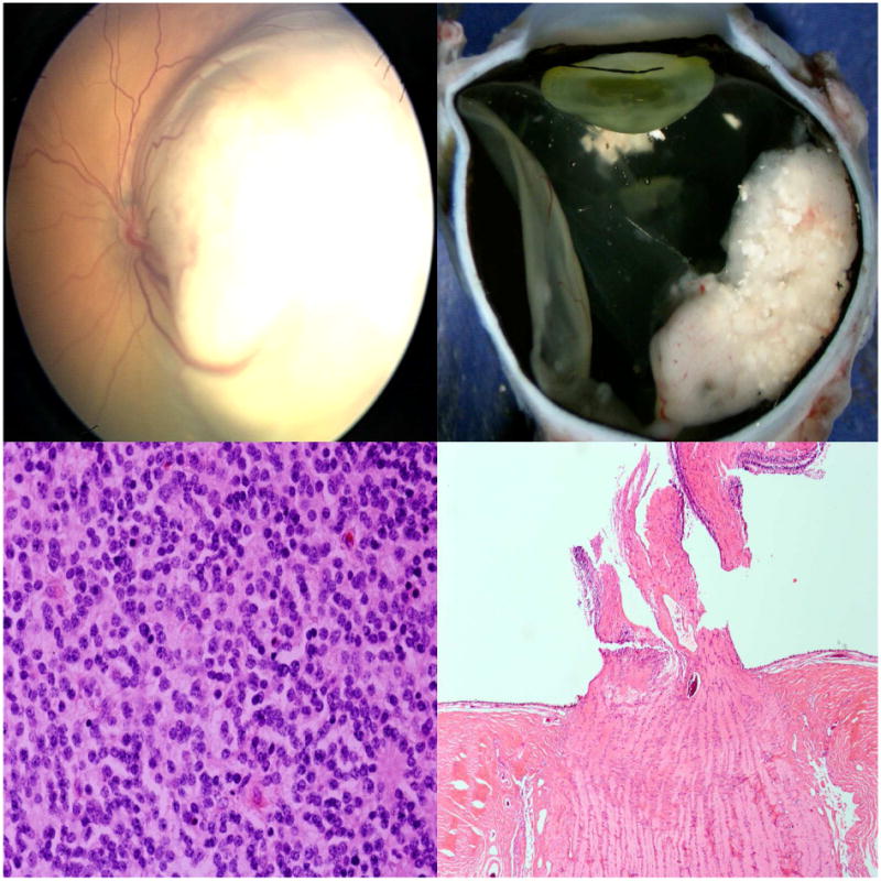
Figure 2.

Mild anaplasia without high-risk features in retinoblastoma. Top left: A whitish, elevated lesion in the left eye involves the macula and temporal retina; Top right: Gross sectioning of the enucleated eye shows a white retinal mass with calcifications; Bottom left: Histologic evaluation reveals mild anaplasia; Bottom right: There is no optic nerve nor choroidal invasion. (hematoxylin and eosin: bottom left, 100×; bottom right, 10×)
