
Figure 1. Comparison of Histological and EM findings in PNPLA8-affected tissues from human and mice.
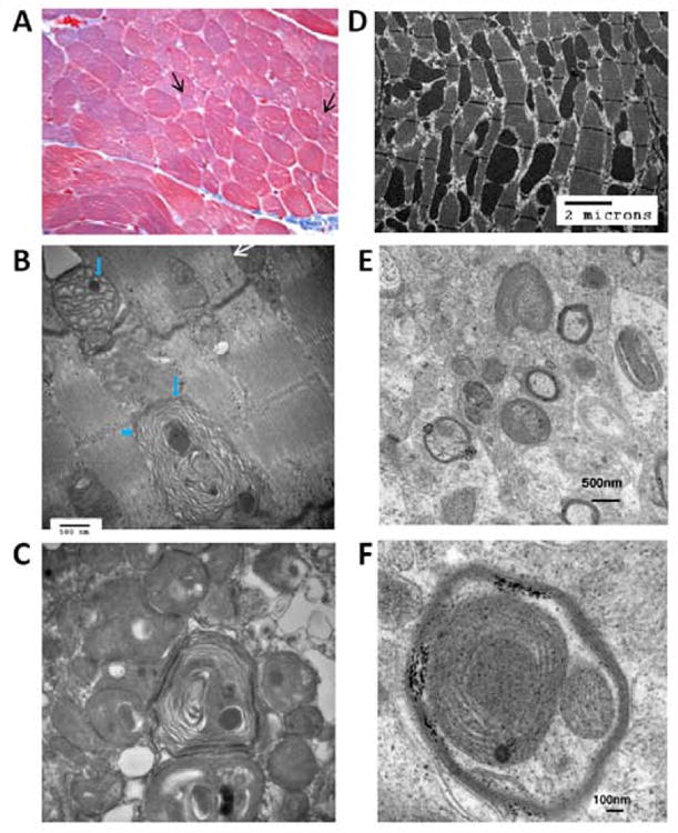
Panels (A)-(C) show paraffin-embedded muscle biopsy from patient CMH193. Trichromestaining (A) (magnification 400 ×) revealed variability in fiber size with isolated smallatrophic fibers (arrows). Normal fibers average 35 microns. ATPase stains (not shown) revealed that atrophic fibers were type 1 and 2 fibers. Ragged red fibers were not found. Oxidative enzyme reactions (SDH and COX) were normal and no large aggregates of mitochondria were demonstrable.
(B) EM photograph of muscle biopsy from CMH193 at 48000 magnification. Mitochondria show abnormal concentric disarray of internal cristae (arrow head) and globular dense osmiophilic inclusions (short arrows). White arrow shows a mitochondrion close to normal. (C) EM photograph of muscle biopsy from CMH193 at 36000 magnification, exhibiting subsarcolemmal aggregates of abnormal mitochondria with concentric lamellar membranes and dense round to oval osmiophilic inclusions. D) Electron micrograph of myocardium from the Pnpla8 -/- mouse; E) Electron micrograph of the cerebellum from Pnpla8 -/- mouse exhibiting dysmorphic mitochondria; F) High power view of the cerebellum from the Pnpla8 -/- mouse.