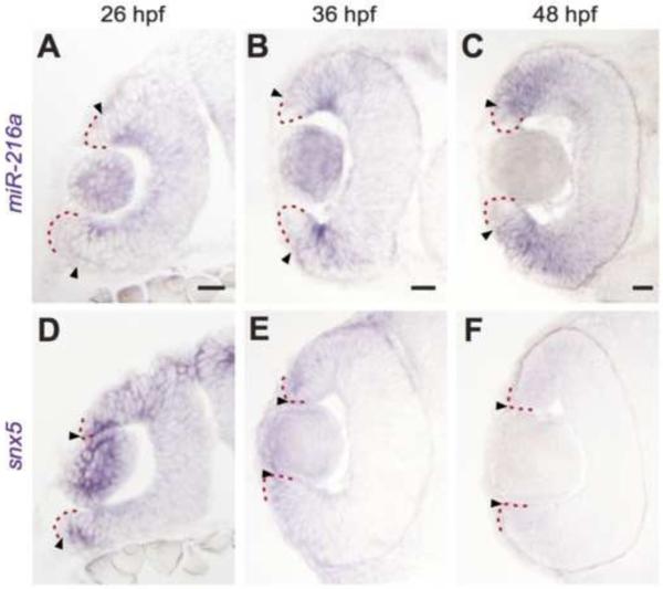
Figure 1. miR-216a and snx5 have complementary expression patterns during development.
Transverse sections of whole mount in situ hybridizations for miR-216a and snx5 at 26 (A,D), 36 (B,E), and 48 (C,F) hours post fertilization (hpf). miR-216a expression spreads from the center of the developing retina toward the periphery. snx5 is detected in a complementary pattern becoming increasingly restricted over time to a small number of cells at the far periphery of the developing retina. Arrowheads indicate the extent of signal, the red dashed line indicates the lateral edge of the optic cup. Scale bar: 20μm.

