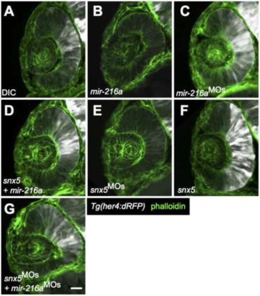
Figure 3. miR-216a and snx5 regulate Notch activation.
Transverse sections of developing retinas from 30 hours post fertilization (hpf) Tg(her4:dRFP) embryos were injected with dye control (DIC; A), miR-216a (B), miR-216aMOs (C), snx5MOs (E), or snx5 mRNA (F). Reporter expression (white) indicates changes in the zone of Notch activation. Partial rescue of Notch activity is shown in (D) and (G) where embryos were co-injected with combinations of either snx5 and miR-216a (D) or snx5MOs and miR-216aMOs (G). Sections were stained with Alexa Fluor 488-conjugated phalloidin (green) to visualize cell boundaries. Scale bar: 20μm.

