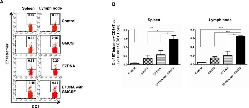
Figure 2. Characterization of E7-specific CD8+ T cells in spleen and lymph node in tumor bearing mice.
C57BL/6 mice (5 per group) were injected with 2 × 10^4 TC-1/luc cells intravaginally then vaccinated intramuscularly with or without 40μg CRT/E7 DNA starting 4 days after tumor challenge for a total of 3 times with 3-day interval. 10 days after tumor challenge, mice were injected with or without 100ng GMCSF intravaginally for 3 times with 2-day interval. 21 days after tumor challenge, spleen and lymph node were harvested and stained with PE-conjugated HPV16 H-2 Db-RAHYNIVTF tetramer and APC-conjugated CD8 monoclonal antibody followed by flow cytometry analysis. (A) Representative flow cytometry showing the percentage of E7-specific CD8+ T cells in spleen and lymph node of various groups. (B) Bar graph depicting the percentage of E7-specific CD8+ T cells in all CD8+ T cells in pooled spleen and lymph node of various groups (mean ± s.d.). * indicates p < 0.05, ** indicates p < 0.01, and *** indicates p < 0.001.

