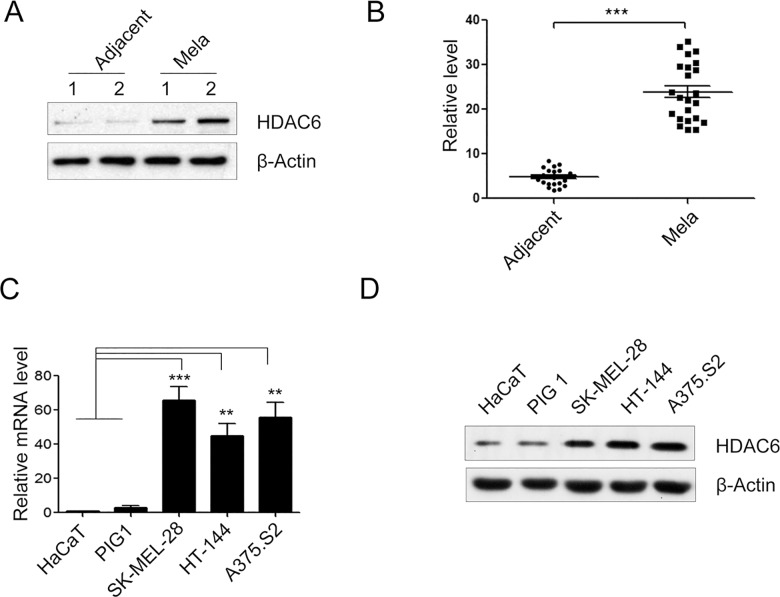
Fig 1. The expression of HDAC6 was up-regulated in melanoma tissues and cell lines.
(A) Representative melanoma tissue (Mela) and adjacent normal dermatic tissue (Adjacent) samples of HDAC6 protein expression were determined by Western blot. β-actin was used as internal loading controls for the cell lysates in the Western blot analysis. (n = 23). (B) The protein expression of HDAC6 in 23 pairs of melanoma tissue (Mela) and adjacent normal dermatic tissue (Adjacent) samples. (C, D) The mRNA level and protein expression of HDAC6 were analyzed in several melanoma cell lines, A375.S2, SK-MEL-28, HT-144 and human immortalised keratinocytes (HaCaT) and normal human epidermal melanocytes (PIG1) by quantitative real time PCR (qRT-PCR) (C) and Western blot (D). (n = 4). Bars represent the mean ± S.E.M. values. Statistical significance (**P < 0.01, ***P < 0.001).

