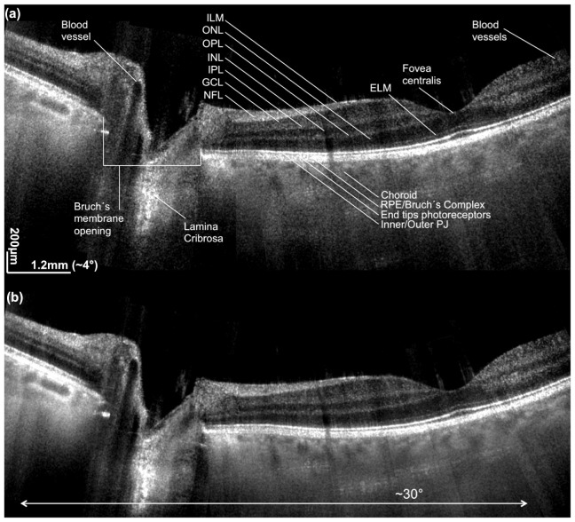
Fig. 4.
Stitched widefield 2D retinal images of macula and optic nerve head (ONH). The total field of view is approx. 30°. (a) is obtained without averaging, (b) is obtained by averaging 4 successive tomograms in scanning direction. ILM is internal limiting membrane, ONL is outer nuclear layer, OPL is outer plexiform layer, INL is inner nuclear layer, IPL is inner plexiform layer, GCL is ganglion cell layer, NFL is nerve fiber layer, ELM is external limiting membrane, RPE is retinal pigment epithelium, PJ is photoreceptor junction.

