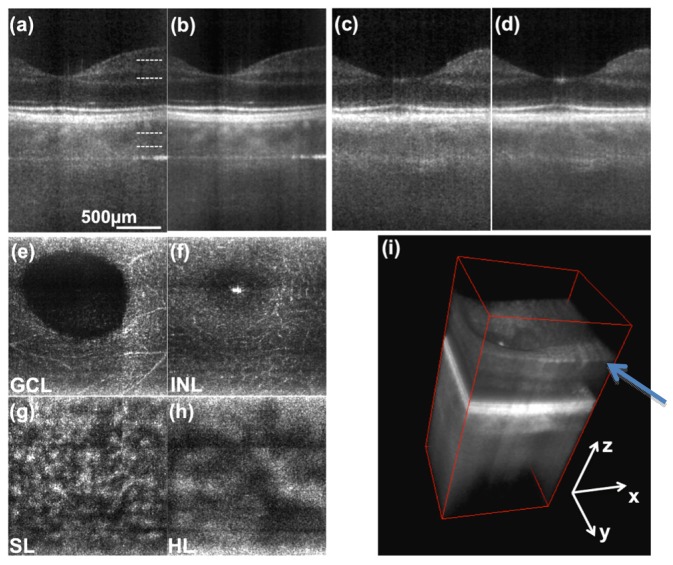
Fig. 5.
3D retinal images of parafoveal region. (a) single frame tomogram in transversal (parallel) direction. (b) averaging 4 successive tomograms in scanning (sagittal) direction. (c) and (d) tomograms along the sagittal coordinate. (e), (f), (g) and (h) are enface projections at depth locations indicated in (a). (i) 3D rendering of same data. The arrow points to visible nerve fiber bundles. (GCL- ganglion cell layer, INL - inner nuclear layer, SL - Sattler’s layer, HL - Haller’s layer).

