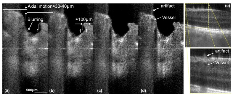
Fig. 11.

Demonstration of the effect of axial motion artifacts. (a)-(d) are tomograms acquired around the ONH with Config. A1. The blurring emanates from eye motion during acquisition of the spectrum. (e) shows a section acquired at approx. 4° eccentricity from the fovea towards the ONH. The artifact highlighted in the magnified image of (e) and in (e) is a consequence of the blurring induced by the axial blood flow component in the vessel below. The distance between location 1 and 1’ indicated in (b) corresponds to the estimated lateral motion between the tomogram in (a) and the successive tomogram in (b). The axial displacement between tomograms in (a) and (b) might be overestimated, since the sample motion introduces a Doppler frequency causing an additional artificial shift of the sample structure.
