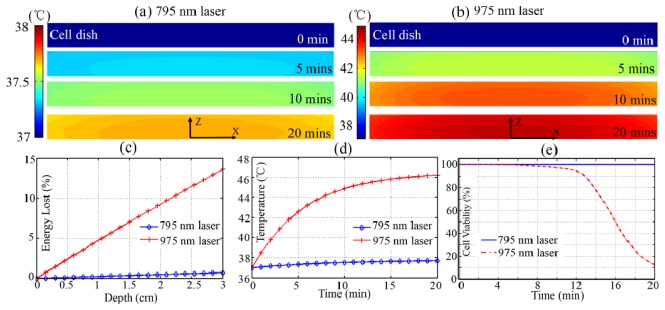
Fig. 3.

Cross-section of the spatiotemporal temperature distributions of cell-in-celldish model under (a) 795 nm and (b) 975 nm laser excitation; (c) The laser energy lost in the 3-mm-thick PBS; (b) Time-resolved temperature variation of adhered cells (0, 0, 0.5 mm) in 20 min; (e) Cell viability prediction after 20 min excitation.
