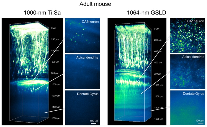
Fig. 3.
Two-photon fluorescence imaging of cortical and hippocampal neurons with 1000-nm MaiTai eHP DeepSee and 1064-nm high-peak power GSLD based light source excitation in adult H-line mouse brain. Maximum intensity projections of three-dimensional stacks were obtained with the 1000-nm TiSa laser (left panel) and 1064-nm GSLD (right panel). Each xy image of the cortical region and hippocampal region was acquired under different scanning conditions. Six normalized xy frames from the z-stack at various depths are shown, including the hippocampal CA1 pyramidal cell layer, apical dendrites, and hippocampal dentate gyrus.

