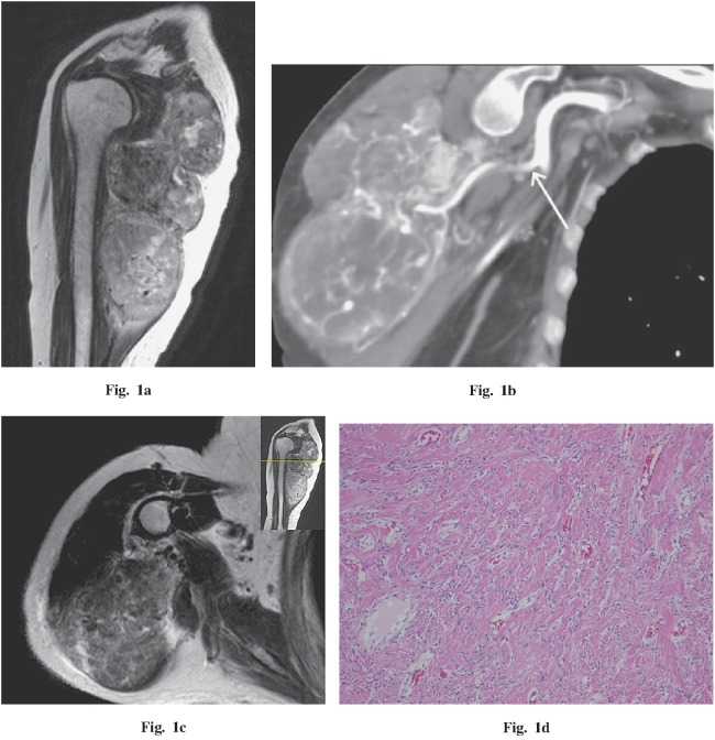
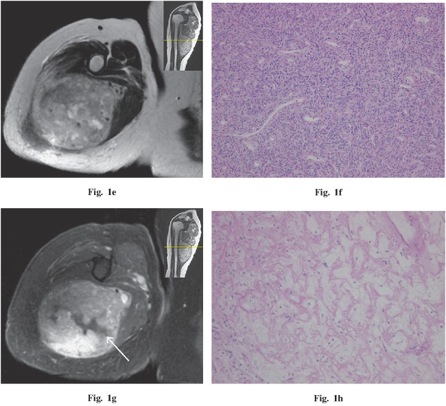
Fig. 1.
A 78-year-old woman with a palpable and painful mass in the right shoulder. a Sagittal T2-weighted image demonstrates a low- to high-intensity, well-circumscribed, lobulated mass compressing the triceps brachii and deltoid muscles. A flow void was detected in the mass. b Coronal oblique CT (arterial phase) demonstrates a vascular pedicle (white arrow) and numerous tumor vessels. c, d Comparing the axial T2-weighted image and histopathology demonstrates that the fibrous area corresponds to the area iso-intense to muscle. e, f Comparing the axial T2-weighted image and histopathology demonstrates that the cellular area corresponds to the moderate to high signal area. g, h Comparing the axial T1 fat-saturated post-gadolinium MR image and histopathology demonstrates that the area of myxoid change corresponds to the area without gadolinium enhancement (g; white arrow). Hematoxylin and eosin stains, 100× magnification.


