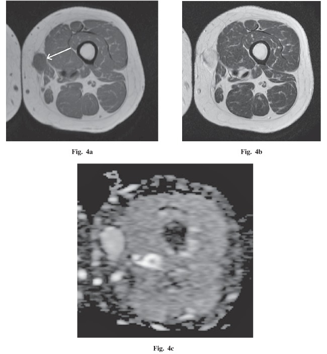
Fig. 4.
A 47-year-old woman with a palpable mass in the left thigh. a An axial T1-weighted image demonstrates an iso-intense, well-circumscribed, oval mass within the subcutaneous tissues adjacent to the medial great and gracilis muscles (white arrow). b An axial T2-weighted image demonstrates the markedly high intensity of this lesion. c The apparent diffusion coefficient (ADC) map demonstrates a homogeneous, high value compared to muscle, with a mean of 1.96×10⁻³ mm2 / second.

