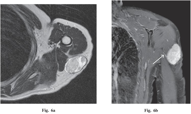
Fig. 6.
A 43-year-old woman with a palpable mass in the left shoulder. a An axial T2-weighted image demonstrates a heterogeneous, high-intensity, well-circumscribed, oval mass within the subcutaneous tissues adjacent to the deltoid and triceps brachii muscles. b Coronal T1 fat-saturated post-gadolinium MR image demonstrates strong enhancement and a small vascular pedicle (white arrow).

